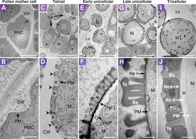
Fig. 2.
Exine development in wild-type Arabidopsis. Transmission electron micrographs showing sections of developing pollen grains in wild-type Arabidopsis (Ler). Micrographs in the top row have the same magnification (A, C, E, G, I) (bars = 10 μm), and those in the bottom row have the same magnification (B, D, F, H, J) (bars = 0.5 μm). (A, B) Pollen mother cells. Each pollen mother cell is surrounded by secondary cell wall. (C, D) Tetrads. Primexine (asterisks) appears between the plasma membrane and callose wall, and the probaculae (arrowheads) are visible above the raised positions of the undulating plasma membrane. Sporopollenin is deposited more densely at the distal part of the probaculae, adjacent to the callose wall. (E, F) Microspores in the early unicellular stage. Feeble tectum, baculae and nexine I are visible. Primexine remains in the cavity of developing exine (asterisks). (G, H) Microspores in the late unicellular stage. Sexine formation is almost complete. (I, J) Tricellular mature pollen grains. Nexine II and intine have formed. Ba, bacula; CW, callose wall; In, intine; Lo, locule; M, microspore; Ne I, nexine I; Ne II, nexine II; PC, pollen coat; PM, plasma membrane; PMC, pollen mother cell; SP, sporopollenin deposited on the outer surface of tetrads; SW, secondary cell wall; T, tapetal cell; Td, tetrad; Te, tectum.

