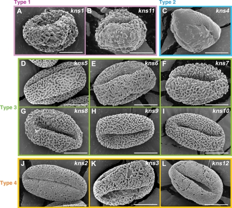
Fig. 3.
Phenotypes of kaonashi mutants and their classification. Scanning electron micrographs of pollen grains isolated from each mutant. (A, B) The type 1 mutants, kns1 and kns11, show highly collapsed exine structure. (C) The type 2 mutant, kns4, has remarkably thin exine. (D–I) The type 3 mutants, kns5, kns6, kns7, kns8, kns9 and kns10, show defects in tectum formation. (J–L) The type 4 mutants, kns2, kns3 and kns12, show abnormal distribution of bacula location. Bars = 10 μm.

