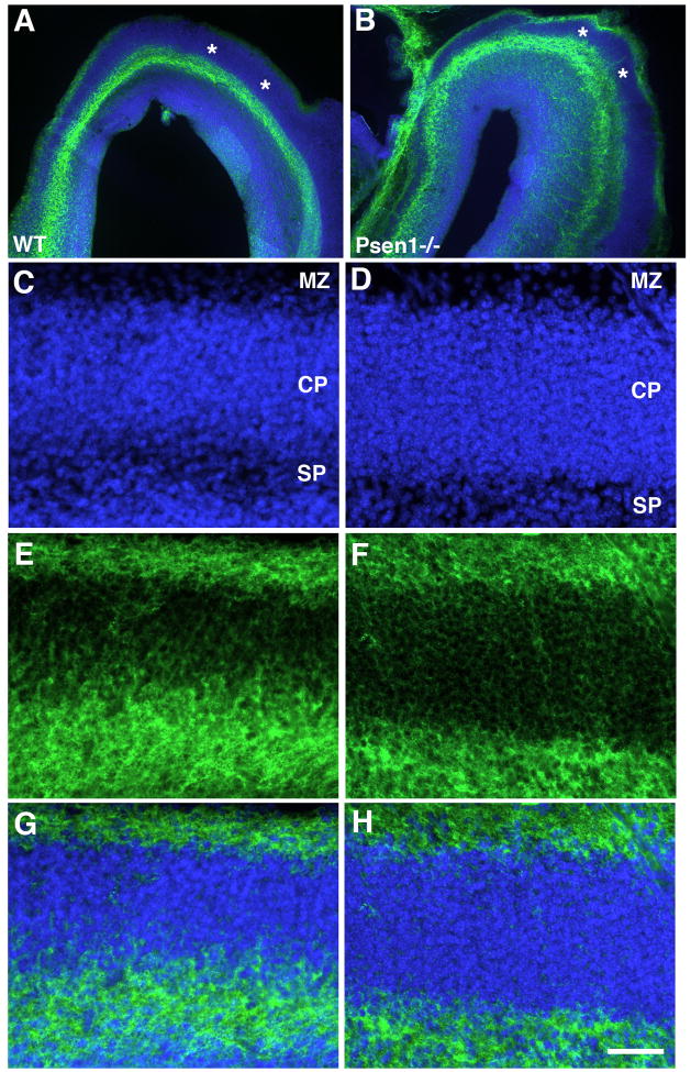
Fig. 5.
CSPG immunostaining reveals splitting of the preplate in Psen1−/− telencephalon. Horizontally cut Vibratome sections from E15.5 wild type or Psen−/− embryos were immunostained with CSPG (green) along with a DAPI nuclear stain (blue). In panels A and B, sections through the frontal poles are shown. Asterisks (*) indicate the unstained cortical plate. In panels C–H, sections from the lateral telencephalon of wild type (C, E, G) or Psen−/− embryos (D, F, H) are shown labeled for DAPI (C, D), immunostained for CSPG (E, F) or as a merged image (G, H) The marginal zone (MZ), cortical plate (CP) and subplate region (SP) are indicated. Scale bar: 200 μm for A–B and 50 μm for C–D.

