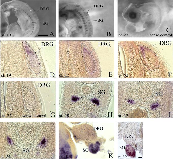
Fig. 1.
Analysis of developmental expression of anaplastic lymphoma kinase (Alk) mRNA in chick peripheral nervous system by whole-mount in situ hybridization. A, B: Transcript is expressed in both sympathetic ganglia (SG) and dorsal root ganglia (DRG) at stages 19 and 23. C: No signal is detected in stage 23 whole-mounts using sense RNA probe as a negative control. D-F: Transverse sections (post-hybridization) reveal Alk expression in the progenitor zones of the DRG at stages 19 (D), 22 (E), and 24 (F). G: Transverse section (post-hybridization) of stage 22 embryo hybridized with sense RNA probe as a negative control. H-L: Transverse sections (post-hybridization) show presence of Alk in SG during stages 19 (H), 22 (I), 24 (J), 35 (K), and 39 (L). Scale bar: A, B, 1 mm; C, 1.5 mm; D, 65 μm; E-J, 120 μm; K, 175 μm; L, 350 μm.

