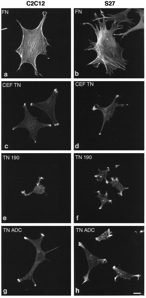
Figure 6.
Confocal microscopy of actin microfilament organization in C2C12 and S27 cells adherent on fibronectin or tenascin-C. Cells were plated onto coverslips coated with 50 nM fibronectin (a and b), 50 nM CEF-TN (c and d), 50 nM recombinant TN-190 (e and f), or 50 nM recombinant TN-ADC (g and h) for 90 min in serum-free medium. After incubation cells were fixed, stained with TRITC-phalloidin, and photographed on a confocal microscope. Bar, 5 μm.

