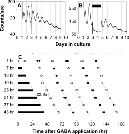
Figure 9. Prolonged Application of GABA Stops the Retinal Clock.
(A and B) Representative retinal explant cultures that received 1 h (A) or 43 h (B) of 3 mM GABA treatment. GABA treatment was started at the beginning of the third circadian cycle. Bars indicate the duration of GABA treatment. Treatment was terminated by fresh media change.
(C) Shown are PER2::LUC expression peaks following GABA washout. Four retinal explants were sampled for each duration of GABA treatment. Bars indicate the duration of GABA treatment. Open circles, filled triangles, open triangles, filled squares, open squares, and filled diamonds indicate first, second, third, fourth, fifth, and sixth peak times, respectively, following GABA washout. Time 0 corresponds to the start of GABA treatment. In the samples with 19–43 h of GABA application, the first peaks appeared ca. 22 h after media change, and the reinitiated rhythms were phase locked to the termination of the GABA pulse, indicating that prolonged application of high dose of GABA stops retinal ensemble PER2::LUC rhythms.

