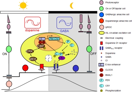
Figure 11. Model for the Circadian Clock Organization in the Mouse Retina.
Dopaminergic amacrine cells (red) and GABAergic cells (blue) reinforce the autonomously generated “day” and “night” states of oscillator cells (yellow) in the inner nuclear layer (INL) through rhythmic secretion of dopamine and GABA, respectively. Dopamine transmission stimulated by excitatory light input from M-cones via ON bipolar cells and/or from intrinsically photosensitive retinal ganglion cells (ipRGCs) likely acts to phase-shift retinal circadian oscillators and reinforce the rising phase of the “day” state of the retinal clock. At night (i.e., molecularly, roughly the falling phase of PER protein rhythms), excitatory input from OFF bipolar cells enhances the activity of GABA, which suppresses rhythmic amplitude of PER oscillations through the GABAA and GABAC receptors and facilitates the molecular resetting effects of dopamine. In addition, endogenous GABA is proposed to reinforce the falling phase of PER rhythms through fostering degradation of accumulated PER protein.

