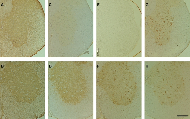
Figure 3. Immunohistochemistry.
Fixed-frozen sections of the spinal cord (10 µm thickness) derived from symptomatic DF mice at 145 days of life (A, C, E, and G) and age-matched NTG mice (B, D, F, and H) were stained with antibodies for ATP1A (A and B), ATP1B (C and D), ATP5A (E and F), ATP5B (G and H). The tissue was counterstained with hematoxylin. Bar: 200 µm.

