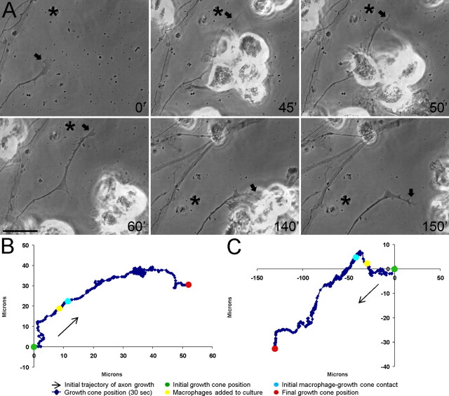
Figure 6.
Macrophages do not induce extensive retraction of actively growing adult dorsal root ganglion axons. A, Six-panel montage of single-frame images from a time-lapse movie in which NR8383 macrophages were added to a culture of actively growing adult dorsal root ganglion neurons on a uniform laminin substrate. Times for each frame are given in the bottom right of each image, and an arrow marks the central domain of the growth cone. An asterisk marks a consistent point in the culture as a reference for growth cone position during frame shifts. Initial macrophage–growth cone contact was made at 45 min (45′). Physical contacts were rapidly broken and no remnant processes were observed between the macrophages and axon at 60′ and 150′. The entire movie can be viewed in supplemental Movie S3 (available at www.jneurosci.org as supplemental material). Scale bar, 20 μm. B, Positional graph tracking the growth cone for entire time-lapse movie. Each point represents the position of the central domain of the growth cone for a single frame (every 30 s). The axon continued to extend ∼40 μm after macrophage contact. C, Positional graph from another representative time-lapse experiment.

