Fig. 3.
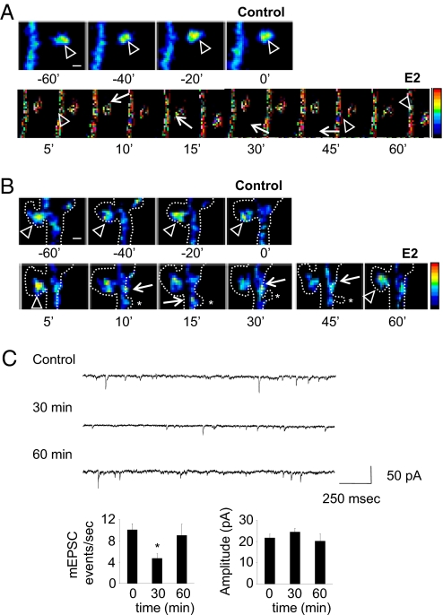
E2 rapidly and transiently induces the formation of silent synapses through trafficking of GluR1 and NR1. (A and B) Time-lapse imaging of neurons expressing GFP-GluR1. Cells were imaged for 60 min before and after administration of E2. Arrowheads indicate GFP-GluR1 in spine heads; arrows indicate GFP-GluR1 in dendritic shaft. Dotted lines indicate neuron outline, as determined by Discosoma red fluorescent protein coexpression; asterisks show transient emergence of novel spines upon E2 treatment. (C) AMPAR mEPSCs after E2 treatment. Frequency and average amplitude of mEPSCs were measured; frequency, but not amplitude, of mEPSCs was significantly reduced at 30 min. *, P < 0.05; ***, P < 0.001. [Scale bars, 1 μm (A and B).]

