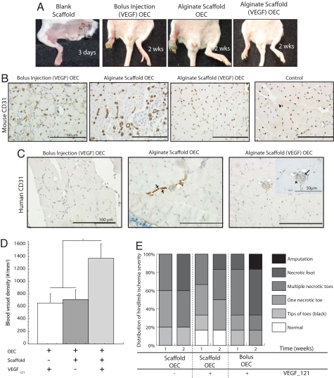
Fig. 2.
Analysis of angiogenesis in ischemic hindlimbs after OEC transplantation. (A) Implantation of blank scaffolds, bolus injection of OECs and VEGF (same quantities as placed in scaffolds), transplantation of OECs on scaffolds lacking VEGF (alginate scaffold OEC), and transplantation of OECs on scaffolds presenting VEGF121 [alginate scaffold (VEGF) OEC]. Photomicrographs of tissue sections from ischemic hindlimbs of SCID mice at postoperative day 15, immunostained for the mouse endothelial cell marker CD-31 (B), and human CD-31 (C). (D) Quantification of the total blood vessel densities in hindlimb muscle tissue after 2 weeks with bolus injection of VEGF121 and OECs (+ − +), scaffold delivery (no VEGF121) of OECs (+ + −), or scaffold delivering OECs with VEGF121 (+ + +) in SCID mice. (E) Hindlimbs subjected to surgery were also visually examined, and grouped as normal (displaying no discrepancy in color or limb integrity from nonischemic hindlimbs of the same animal), or presenting one necrotic toe, multiple necrotic toes, or a complete necrotic foot. Mean values are presented with standard deviations (n = 6) in both graphs. *, P < 0.05 between conditions.

