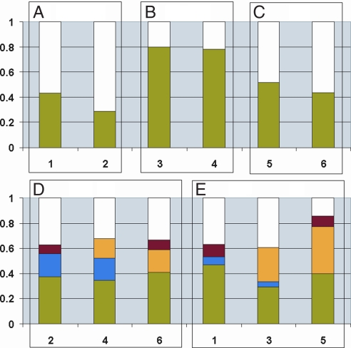
Fig. 3.
Comparison of the overlap in the protein coronas of the different polystyrene particles. Graphs: 1, 100-nm amine-modified; 2, 50-nm amine-modified; 3, 100-nm plain; 4, 50-nm plain; 5, 100-nm carboxyl-modified; and 6, 50-nm carboxyl-modified. The fractions shown are calculated without including different Ig chains. (A–C) Comparison of the similarity between the coronas around different size particles with similar surface properties: fraction of proteins found on both particles (green), and fraction of proteins that is found on one size but not the other (white). (D and E) Comparison of the similarities of the corona for particles of the same size but different surface charges: fraction of proteins found on all three particles (green), fraction of proteins found on the amine-modified and plain particles (blue), fraction of proteins found on the plain and carboxyl-modified particles (yellow), fraction of proteins found on the amine- and carboxyl-modified particles (red), and fraction of proteins found on just one specific particle surface (white).

