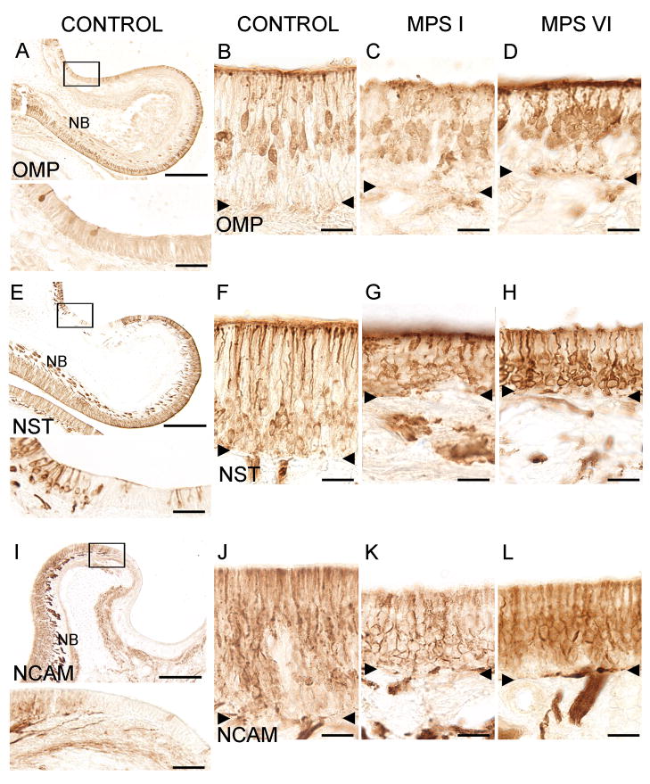
Figure 2.
Immunohistochemical localization of ORNs in control and affected cats. A. OMP-immunoreactivity (ir) of mature ORNs in a region of the ethmoturbinate in a control animal. Inset: a higher magnification of the boxed area shows individual labeled cells and no labeling of adjacent respiratory cells. B. OMP-ir cells project dendritic processes to the apical surface of the sensory epithelium with their cell bodies localized in the middle layers of the tissue. C. OMP-ir was detected in MPS I-affected animals. D. OMP-ir was detected in MPS VI-affected animals. E. NST-ir in olfactory epithelium of a control cat. Cells are labeled in the epithelium and NST-ir olfactory nerve bundles are seen below the basement membrane in the lamina propria. Inset: Higher magnification of NST-ir in ORNs. Left portion of the epithelium contains NST-positive cells while center portion shows non-sensory epithelium, which lacks NST-ir cells. F. NST-ir cell bodies tend to be found above the basement membrane and below the OMP positive cells (2B) suggesting that NST labeled more immature ORNs. Strong ir was also present in the dendrites and olfactory knobs. G. NST-ir ORNs were present in MPS I animals. H. NST-ir ORNs were also present in MPS VI animals. I. NCAM-ir in olfactory epithelium from a control cat. NCAM-ir (sensory) and NCAM negative (non-sensory) epithelium is visible. Note the pronounced labeling of nerve bundles below the sensory portion of epithelium (NB). Inset: Higher magnification of the transition area between sensory (left) and non-sensory epithelium (right) which is clearly demarcated by NCAM-ir. J. NCAM-labeled cells throughout the sensory part of the epithelium in control cats. K. NCAM –ir was detected in the epithelium of MPS I animals. L. NCAM-ir was detected in MPS VI animals. The reduced OE thickness in both MPS I and MPS VI animals can be seen in their respective images. NB = nerve bundles. Arrowheads = basement membrane. Scale bars are 250 μm for A, E, and I; 40 μm for the insets and 20 μm for B–D, F–H and J–L.

