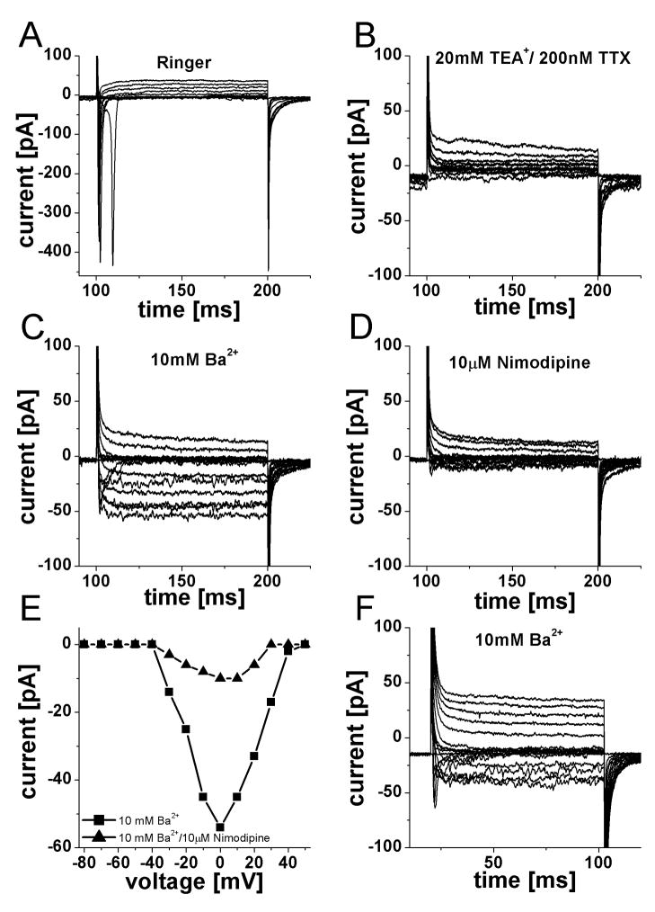
Figure 6.
Voltage-activated calcium currents were present in ORNs from both control and MPS I-affected cats. A. Family of current elicited by pulse protocol from Fig 5B, in Ringer solution, a fast sodium inward current was observed. B. Addition of 20 mM tetraethylammonium+ (TEA+) and 200nM tetrodotoxin (TTX) reduced the outward currents and abolished the sodium current. C. Further addition of 10 mM Ba2+ enhanced voltage-activated calcium currents present in this cell. A fast, transient component as well as a sustained current is visible. D. Adding 10 μM Nimodipine (a potent L-type calcium channel blocker) greatly reduced both components of inward current. E. Current-voltage relationship for the sustained inward current in the presence of 10 mM Ba2+ and after addition of 10 μM nimodipine. Nimodipine reduced the current amplitude by 81%. F. Recording from an ORN from a control cat under identical conditions as in C. Two inward current components of comparable size and time course were observed.

