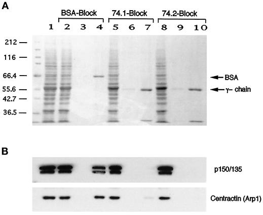
Figure 5.
Monoclonal antibodies 74-1 and 74-2 block dynein IC–dynactin interaction in vitro. Rat brain cytosol (lane 1) was loaded onto a dynein IC affinity column (see MATERIALS AND METHODS) that was pretreated with BSA (lanes 2–4), m74–1 (lanes 5–7), or m74–2 (lanes 8–10). The columns were extensively washed and eluted with 1 M NaCl. The fractions were resolved by SDS-PAGE followed by transfer onto Immobilon and stained with Coomassie brilliant blue (A) and subsequently probed with antibodies to p150Glued and centractin (B). Lane 1, cytosol; lanes 2, 5, and 8, flow-through fractions; lanes 3, 6, and 9, final wash fractions; lanes 4, 7, and 10, fractions eluted with 1 M NaCl.

