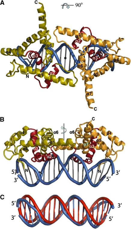
Figure 3.
Structure of the LFY-C dimer bound to DNA. (A, B) Two orthogonal views of the LFY-C dimer (residues 237–399) bound to DNA. Monomers are coloured in olive and orange with the helix-turn-helix (HTH, helices α2 and α3) motif in red. The DNA duplex is depicted in blue. Figures 3, 4B, 5A and 6 were produced with program Pymol (Delano, 2002). (C) Superposition of the DNA duplex found in the LEAFY–DNA complex (blue) with regular B-form DNA (red).

