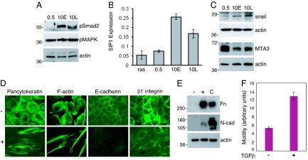
Fig. 2.
TGFβ signaling is activated in vHMEC-ras cells with a mesenchymal morphology and can induce EMT in vHMEC-ras cells with an epithelial morphology. Immunoblot analysis of phospho-Smad2 and phospho-MAP kinase (A) or Snail and MTA3 (C) on cell lysates prepared from vHMEC-ras0.5 (0.5) and vHMEC-ras10 early- (10E) and late- (10L) passage cells. (B) Quantitative real-time PCR (qPCR) analysis of SIP1 expression. (D) Immunofluorescence analysis of vHMEC-ras0.5 untreated (−) or treated (+) with 2 ng/ml TGFβ for 72 h and immunostained as indicated. (E) Immunoblot analysis of fibronectin (Fn) and N-cadherin (N-cad) on cell lysates prepared from vHMEC-ras0.5 either untreated (−) or treated (+) as in (D). Cell lysates prepared from fibroblasts were used as a positive control (C). (F) Transwell motility assay depicting vHMEC-ras0.5 cell migration toward media not supplemented (−) or supplemented (+) with 10 ng/ml TGFβ as a chemoattractant for 48 h.

