Fig. 4.
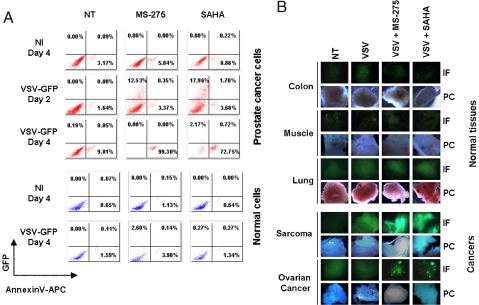
HDI pretreatment enhances VSV oncolytic activity in primary tumor specimens. (A) Ex-vivo primary cancer or normal prostate cells were subjected to 24 h HDI pretreatment followed by VSV-Δ51GFP infection (5 MOI). VSV replication (GFP, y axis) and apoptosis induction (AnnexinV-APC staining, x axis) were determined at 2 and 4 days after infection by FACS. NT denotes non-HDI treated cells, NI denotes non-VSV-infected samples. (B) Human ex-vivo cancer or normal tissue specimens were inoculated with VSV-Δ51-GFP in the absence or presence of HDI pretreatment for 7 h. GFP expression was monitored 48 h after viral inoculation by fluorescence microscopy (IF). Phase contrast (PC) images of tissue samples are shown. NT denotes non-HDI treated, non-VSV-infected cells.

