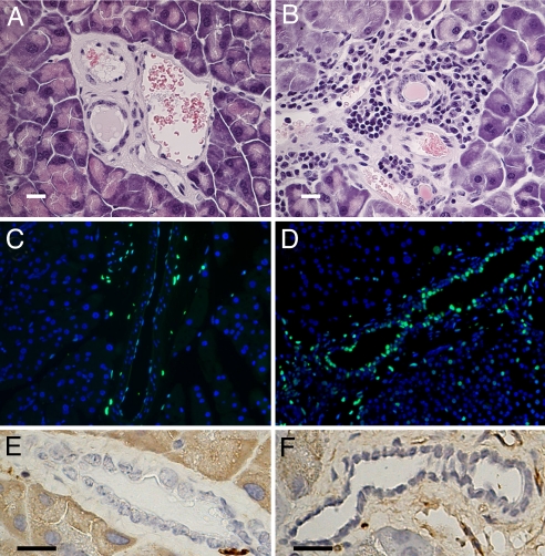
Fig. 2.
Immuno-morphological study of 11-month-old female pancreas. (A and B) Hematoxylin-eosin staining demonstrates a large infiltration of immune cells all around medium-sized pancreatic ducts of LXRβ−/− mice (B) compared with WT mice (A). (C and D) TUNEL staining (green) shows more apoptotic epithelial cells in the pancreatic ducts in LXRβ−/− mice (D) than in WT mice (C). Nuclei are counterstained with DAPI. (E and F) There are no detectable differences between WT mice (E) and LXRβ−/− mice (F) in BrdU-positive cells in pancreatic ducts. (Scale bars: 10 μm.)

