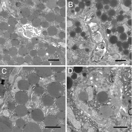
Fig. 3.
Pancreatic electron microscopy study of 11-month-old male mice. (A and B) Laminar structures, characteristic of sticky secretions in luminal region of pancreatic ducts, are detectable in LXRβ−/− mice (B) where a dilatation of the ducts is also evident compared with WT mice (A). (C and D) Cisternae of Golgi apparatus are more dilated in LXRβ−/− mice (D) than in WT mice (C). (Scale bars: 1 μm.)

