Fig. 5.
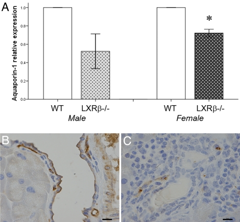
Expression of AQP1 in pancreas of WT and LXRβ−/− mice. (A) mRNA levels of AQP-1 are significantly decreased in 11-month-old LXRβ−/− female mice. Values of WT mice were taken as 1, and the knockout mouse values are expressed relative to WT mice. Data represent the mean ± SE. *, P < 0.05 versus WT. (B and C) Protein levels detected with immunohistochemistry are markedly reduced in pancreatic ducts of LXRβ−/− female mice (C) compared with WT mice (B). Positive staining is detectable on the luminal membrane of ductal epithelial cells in WT mice. Staining of AQP-1 in endothelial cells of small blood vessels is still detectable in the knockout animal. (Scale bars: 10 μm.)

