Figure 2.
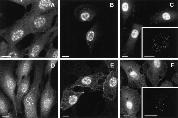
Indirect immunofluorescence of NRK-49F rat fibroblasts indicated nuclear localization of PIPKIα (A–C) and PIPKIIα (D–F). The speckled staining in nuclei was observed with methanol fixation (A and D), or with a brief preextraction using 0.2% Triton X-100 and then fixation with formaldehyde (B and E). Strong diffuse nuclear staining upon formaldehyde fixation was obtained when anti-PIPK antibodies were used at 10 μg/ml (C and F). However, this picture at lower concentrations progressed to speckles and then resolved into a pattern of smaller dots when 0.5 μg/ml anti-PIPKIα or 1 μg/ml anti-PIPKIIα antibodies were used (C and F, insets). Insets, thin optical sections of magnified view of the nuclei of human MG-63 cells. Fixation and staining protocols are detailed in MATERIALS AND METHODS. Bar, 10 μm.

