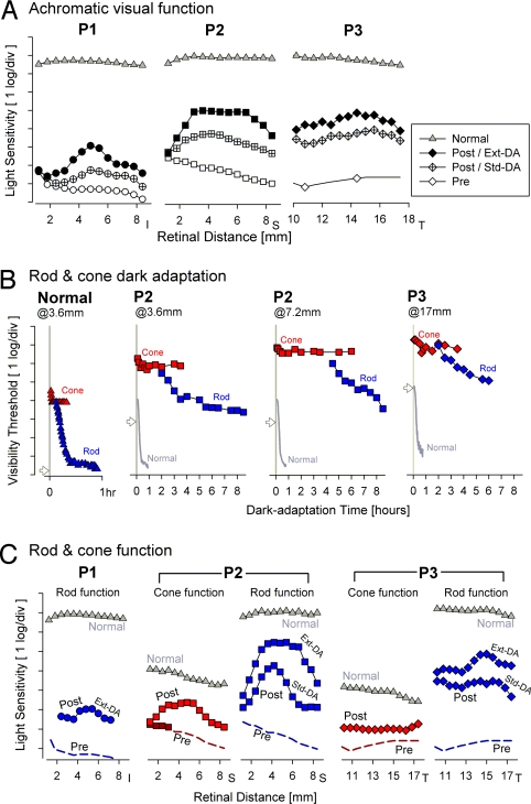
Fig. 3.
Rod- and cone-photoreceptor-mediated visual function across the retinal region of study eyes showing biological activity. (A) The magnitude of the biological activity can be improved by allowing for a period of extended dark adaptation (Ext DA) before testing. Changes in the shapes of the light sensitivity profiles in patient 1 and patient 2 between standard dark adaptation (StdDA) and extended dark adaptation conditions suggest local differences in adaptation rate. (B) Dark adaptation kinetics measured with chromatic stimuli after a 7 log scot-td.s yellow adapting flash (presented at time 0) in patient 2 (at 3.6 and 7.2 mm inferior loci) and patient 3 (at 17 mm temporal locus). Also shown are detailed results from one normal (N) subject at 3.6 mm inferior (Left) and mean results from normal subjects at each location (gray lines). Cone adaptation kinetics (red symbols) are fast and do not show a difference from healthy cones. Rod adaptation kinetics (blue symbols) are extremely slow compared with healthy rods, and in patient 2 there is evidence for a large intraretinal difference in recovery rate. Additionally, absolute thresholds of both rod and cone systems are abnormally elevated. Note, the vertical threshold scale is inverted compared to the sensitivity scale in A to be consistent with traditional presentations of such data sets. (C) Maximal improvement in rod-mediated function (blue) across the region of treated retina ranged from 2.3 log in patient 1, 4.8 log in patient 2, and 4.5 log in patient 3; cone vision improvements (red) range from 1.7 log in patient 2 to 1.2 log in patient 3. Note that pretreatment estimates for rod- or cone-mediated vision (dashed lines) are best-case extrapolations from achromatic sensitivity measures. Only in patient 2 were pretreatment chromatic sensitivities measurable at 1–3 mm superior retina, and these measures were consistent with cone mediation. For patient 1, after treatment, specialized testing with a 4° diameter stimulus centered at 4.8 mm inferior retina suggested rod mediation for the blue stimulus. Pretreatment (Pre) results are from ≈18 to ≈3 months before treatment; posttreatment (Post) results are from 1–3 months after treatment.

