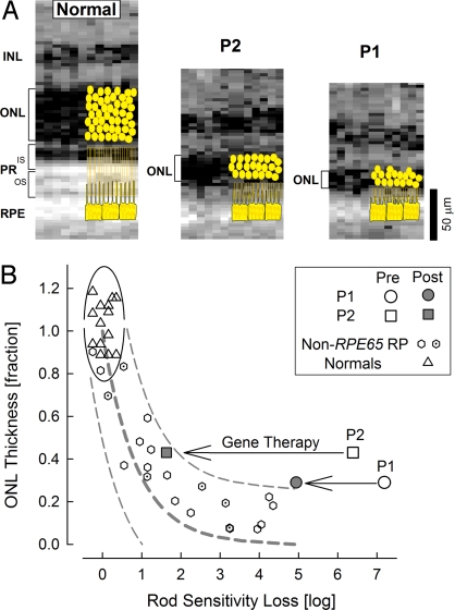
Fig. 4.
Pathophysiology of complex visual loss resulting from photoreceptor degeneration and biochemical blockade, and the effect of gene therapy. (A) Retinal cross-sectional images at the region of maximal biological activity show the photoreceptor layer (ONL, brackets) to be abnormally thinned; greater degeneration of photoreceptors is evident in patient 1 compared with patient 2. Normal retina and patient 2 are at ≈5 mm superior retina, patient 1 is at ≈5 mm inferior retina. Schematics of photoreceptors and RPE are overlaid (yellow) and to the right of the actual optical images; these are derived from cynomolgus monkey histology (3, 28). INL, inner nuclear layer; PR-IS and PR-OS, photoreceptor inner and outer segments. (B) The relationship between photoreceptor layer thickness and rod sensitivity loss in patients 1 and 2 before and after therapy compared with normal subjects and patients with retinitis pigmentosa without mutations in the RPE65 gene. Normal variability is described by the ellipse encircling the 95% C.I. of a bivariate Gaussian distribution. Dotted lines define the idealized model of the relationship between structure and function in pure photoreceptor degenerations and the region of uncertainty that results from translating the normal variability along the idealized model. Gene therapy (arrows) results in partial (patient 1) or a nearly complete (patient 2) amelioration of the biochemical blockade transforming the complex RPE65-LCA disease phenotype into a simpler retinal degeneration phenotype.

