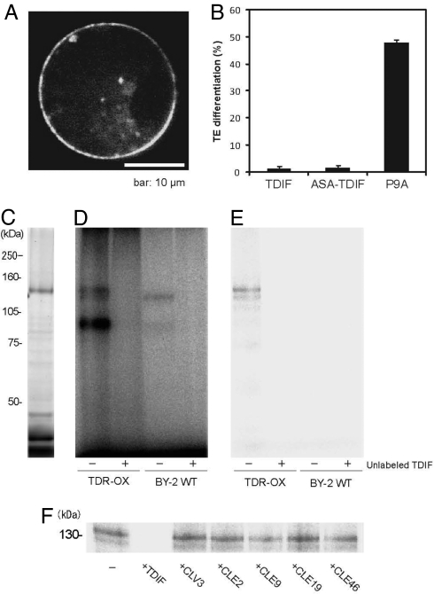
Fig. 3.
Direct interaction between TDR and TDIF. (A) Subcellular localization of TDR. (B) Suppression of TE differentiation in Zinnia cell culture. TDIF, ASA-TDIF, and the nonfunctional peptide P9A are shown. (C) Visualization of TDR-ΔKD-HT in transgenic tobacco BY-2 cells overexpressing TDR-ΔKD-HT. (D) Photoaffinity labeling of TDR-ΔKD-HT by 125I-ASA-TDIF in the absence (−) or presence (+) of excess unlabeled TDIF in microsomal fractions from TDR-ΔKD-HT-overexpressing cells (TDR-OX) or nontransformed tobacco BY2 cells (BY2-WT). (E) Immunoprecipitation with an anti-HaloTag antibody for the photoaffinity-labeled samples used in D. (F) Photoaffinity labeling of TDR-ΔKD-HT by 125I-ASA-TDIF in the absence (−) or presence of excess unlabeled TDIF, CLV3, CLE2, CLE9, CLE19, or CLE46 peptide.

