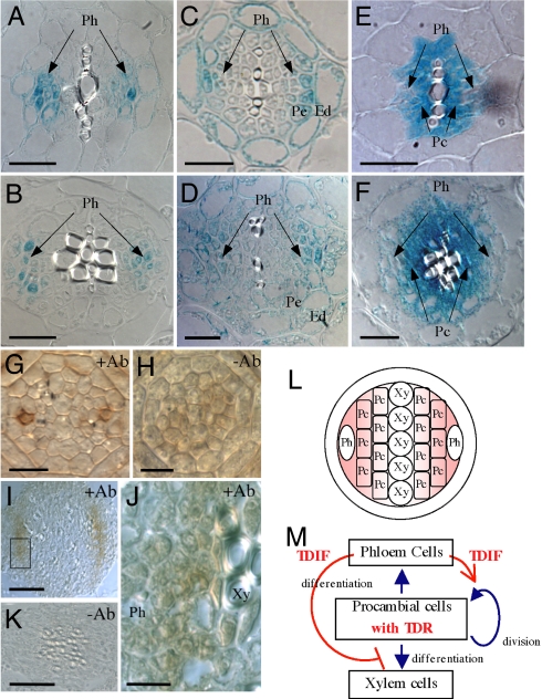
Fig. 4.
Vascular cell-specific localization of TDIF and TDR. (A–F) Distinctive vascular cell-specific expression of the CLE41, CLE44, and TDR genes in roots (A, C, and E) and hypocotyls (B, D, and F). (A and B) pCLE41::GUS. (C and D) pCLE44::GUS. (E and F) pTDR::GUS. Ph, phloem; Pc, procambium; Pe, pericycle; En, endodermis. (G–K) Immunohistochemical localization of the TDIF peptide in the root tip (G and H) and hypocotyls (I–K). (J) Magnification of the box in I. Note that the signal is detected around the phloem cells. (L and M) Positional (L) and functional (M) models of the TDIF/TDR signaling system, which regulates the fate of procambial cells in a non-cell-autonomous manner. (Scale bars: A–F, 20 μm; G and H, 10 μm; I and K, 50 μm; J, 10 μm.)

