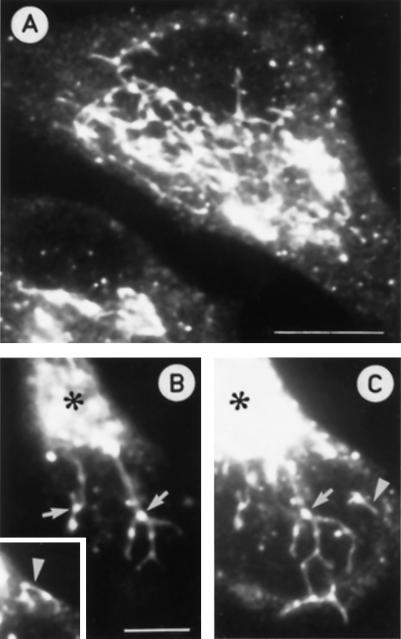
Figure 5.
Visualization of the pre-Golgi tubules in Baf A1-treated PC12 cells using antibodies against rab1p. (A) Confocal immunofluorescence image demonstrating tubulation of the pre-Golgi structures in the drug-treated cells and the formation of an extensive, partly continuous reticulum. (B and C) Two partly overlapping confocal sections from the same cell showing details of a rab1p-positive reticulum that extends from the Golgi region (asterisks) towards the cell periphery. Note the localization of the globular IC domains to the branchpoints of the reticulum (arrows). The arrowheads indicate rab1p-positive tubules extending from and connecting the peripheral, globular pre-Golgi structures. Bars, 10 μm (A) and 5 μm (B).

