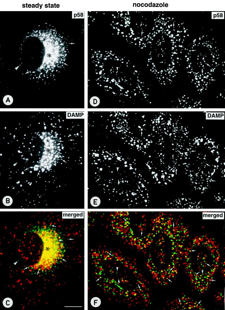
Figure 8.
The lumenal pH of centrally located pre-Golgi structures is acidic. (A–C) NRK cells were incubated for 30 min at 37°C in medium containing the weak base DAMP (50 μM), followed by fixation and double-staining for p58 (A) and DAMP (B). C is the merged image, showing apparent colocalization of the two markers in the perinuclear Golgi region (asterisks). Some colocalization is also seen in a few more peripherally located pre-Golgi structures that can be distinguished as individual elements (arrows). (D–F) To disperse both endocytic and pre-Golgi structures, cells were treated for 2 h with 5 μM nocodazole, incubated in the presence of DAMP, and processed for confocal immunofluorescence microscopy as above. A number of the p58-positive structures colocalize with DAMP in these cells (arrows). The arrowheads indicate pre-Golgi structures that contain very weak DAMP labeling, giving rise to a light green color (instead of yellow) in the merged image. Bar, 10 μm.

