Figure 9.
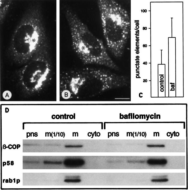
β-COP is redistributed in response to Baf A1 treatment, but its membrane-bound pool is not significantly affected. (A and B) Confocal immunofluorescence images from control (A) and Baf A1-treated NRK cells (B), stained with antibodies against β-COP, demonstrating an increased association of β-COP with peripheral pre-Golgi structures in response to Baf A1. Bar, 10 μm. The quantitation in C shows that Baf A1 causes an approximately twofold increase in the number of detectable pre-Golgi structures. (D) Association of β-COP with membranes. Postnuclear supernatant (pns), total membrane (m), and cytosol (cyto) fractions were prepared from control and Baf A1-treated PC12 cells. Equal amounts of protein from each fraction were run in SDS-PAGE, transferred to nitrocellulose, and analyzed for their content of β-COP, p58, and rab1p by quantitative immunoblotting.

