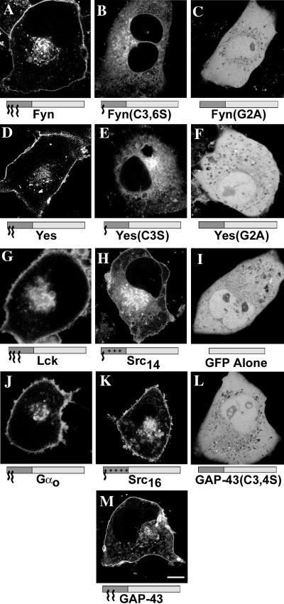
Figure 3.
Subcellular localization of various N-terminal fatty acylated GFP chimeras and mutants in living COS-7 cells. GFP fluorescence of COS-7 cells expressing FynGFP (A), Fyn(C3,6S)GFP (B), Fyn(G2A)GFP (C), YesGFP (D), Yes(C3S)GFP (E), Yes(G2A)GFP (F), LckGFP (G), Src14GFP (H), GFP alone (I), GαoGFP (J), Src16GFP (K), GAP43(C3,4S)GFP (L), and GAP-43GFP (M). Intrinsic GFP fluorescence was detected by confocal laser scanning microscopy. In the models below the images, the dark gray box indicates the appended acylation amino acid sequence and the light gray box indicates GFP. Small acyl chain (∼), myristate; large acyl chain (∧∧∧), palmitate; plus (+) signs, polybasic region. Bar, 10 μm.

