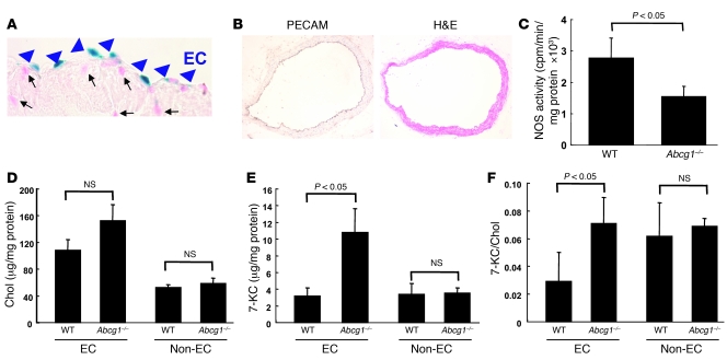
Figure 4. LacZ expression in Abcg1–/– mice and NOS activity and sterol mass in WTD-fed WT and Abcg1–/– mice.
(A) LacZ expression in endothelium of aorta in Abcg1–/– mouse. Blue nuclear lacZ expression was detected specifically in ECs (arrowheads) but not in other cells indicated by nuclear fast red (arrows). (B–F) WT and Abcg1–/– mice were put on a WTD for 12 weeks (n = 4 per group). (B) PECAM-immunostained aorta. Original magnification, ×200 (A), ×100 (B). (C) Aortic NOS activity. (D) Cholesterol mass, (E) 7-KC mass, and (F) 7-KC/cholesterol ratio in ECs and non-ECs from mouse aortas. The results are represented as mean ± SEM.

