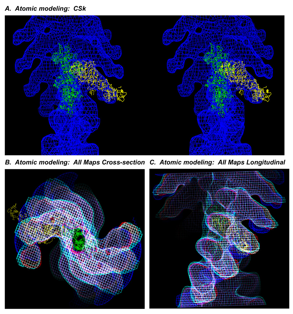
Figure 4.
(A) Nucleotide-free, chicken skeletal S1 crystal structure 22 (yellow) docked on actin 23 (green) modeled into the acto-CSk S1 map (blue, wireframe) in stereo, longitudinal view. Stereo image was prepared using the UCSF Chimera package from the Resource for Biocomputing, Visualization, and Informatics at the University of California, San Francisco. The RLC region of the crystal structure protrudes from the S1 envelope. (B) Cross-section and (C) longitudinal views of all of the density maps in wire frame superimposed, CSk (blue), wt Dros (red), Leth (cyan), IFI-EC (white), and compared to the nucleotide-free, chicken crystal structure (yellow) docked on actin (green) in the actoS1 reconstructions. Truncation of the insect maps after the ELC emphasizes that the ELC of the crystal structure is veering off the straight path of the IFM envelope toward the bend of the chicken RLC.

