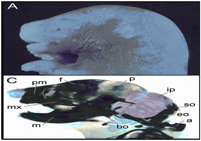
Figure 1.
Fetal heads at gestational day 17 demonstrating characteristic lesions associated with valproic acid treatment. A, Control; B, Exencephaly and open eye. These heads were then cleared and stained with alcian blue for cartilage and alizarin red for bone. C, Control; D, Exencephalic fetus demonstrating abnormalities in most of the craniofacial skeleton. The following structures are labeled: m, mandible; mx, maxilla; pm, premaxilla; f, frontal; p, parietal; ip, interparietal; so, supraoccipital; eo, exoccipital; bo, basioccipital; a, atlas.

