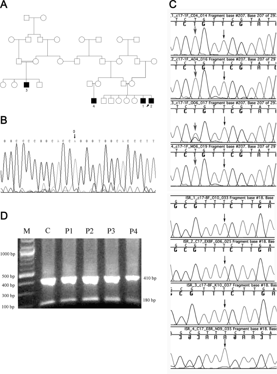
Figure 1.
Genetics of the affected kindred. A, Family tree showing the relationships among the four patients studied. The affected individuals are numbered as in the text. B, Sequence analysis of the POR gene; the mutation G→A at position 1697 in exon 12 causing the replacement of G539R is marked by an arrow on the sequence trace of patient 2. This mutation was identified in all four patients. C, Sequence analysis of CYP17A1. Upper panel (four tracings), Exon 1. Black arrows indicate that the previously reported T deletion at position 198 in CYP17A1 exon 1 was not presented in all four patients. Gray arrows indicate that patients 1, 2, and 4 are homozygous for the polymorphism 195 G→T, and patient 3 is heterozygous. Lower panel (four tracings), Exon 8. Black arrows indicate that the substitution 1235 T→G, reportedly responsible for the missense mutation F147C, was not present in all four patients. D, Digestion of CYP17A1 exon 8 PCR product with Hpy188III. M, Marker 100 bp (New England Biolabs); C, nlormal control; P1–P4: patients 1–4. The 591-bp PCR product of CYP17A1 exon 8 was cut into fragments of 181 and 410 bp in all four patients and in the normal control, confirming the presence of the wild-type sequence.

