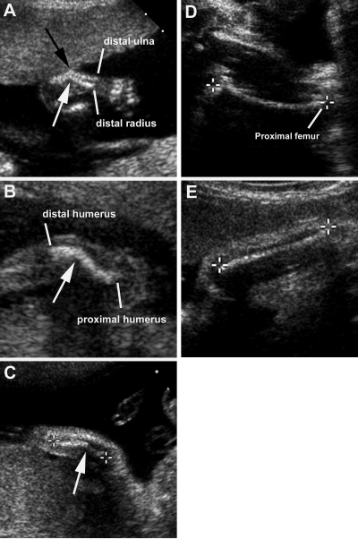
Figure 1.
Sonographic images of sib no. 1. A, Sharp mid-diaphyseal angulation of his right radius (white arrow) and ulna (black arrow) at 18-wk gestation. B, Sharp, mid-diaphyseal angulation (arrow) of his right humerus at 20-wk gestation. C, Improvement in angulation (arrow) of his right radius at 26-wk gestation. Calipers mark the ends of the radius. D, Mildly curved left femur at 34-wk gestation. Calipers mark the ends of the femur. E, Improvement over time of angulation of his right radius at 34-wk gestation. Calipers mark the ends of the radius.

