
Figure 5.
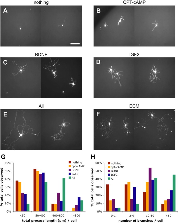
Morphology of CTB-immunopurified CSMNs in vitro. A–F, Representative images of purified P3 CSMNs cultured for 5 DIV in basal serum-free medium (A), basal media containing CPT-cAMP alone (B), CPT-cAMP plus BDNF (C), IGF2 (D), BDNF plus CXCL12 plus IGF2 plus PTN (E), or 10× ECM (F). All live cells were visualized by incubation in calcein AM. Scale bar, 100 μm. G, H, Histograms showing percentages of purified P3 CSMNs alive at 5 DIV in indicated media, visualized by calcein AM incubation, demonstrating <50, 50–400, 400–800, or >800 μm total process outgrowth (G), or 0–1, 2–9, 10–50, or >50 total process branch points (H). In each condition, >20 randomly selected live cells were analyzed.