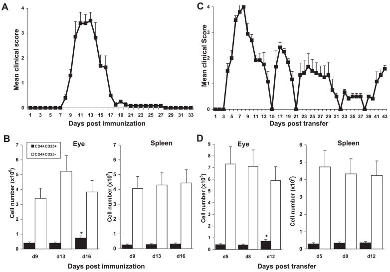
Figure 1.
Proportions of CD4+ cells expressing CD25 in the eye, but not the spleen, increased during recovery in m-EAU and r-EAU. m-EAU (A) or r-EAU (C) was induced and disease severity was determined (n = 10 rats). The mean score at each time point is the average clinical score of 10 independent scores. Eyes and spleens of m-EAU (B) or r-EAU (D) rats were taken at the times indicated (4 rats/day), and the numbers of CD4+CD25+ and CD4+CD25+ cells in the eye and spleen were assessed by FACS staining. Data are expressed as mean ± SD of four individual rats per time point, representative of three separate experiments. Statistical analyses were performed using one-way ANOVA with Tukey post hoc analysis. CD4+CD25+ cell number in the recovery phase was significantly different from that at the onset or peak of disease (*P < 0.01).

