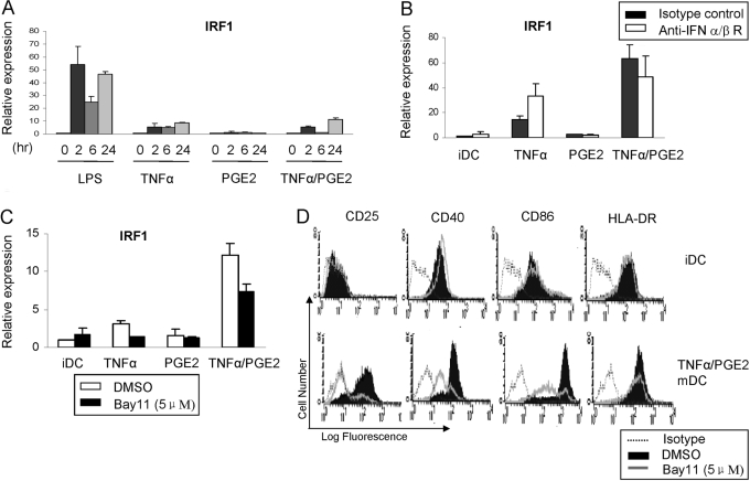
Fig. 4.
TNF-α + PGE2-induced IRF1 expression is partially dependent on NF-κB but not on IFN-α/β. (A) mRNA levels of IRF1 in immature DC treated with LPS, TNF-α, PGE2, or TNF-α + PGE2 for indicated times were measured by real-time PCR and normalized relative to GAPDH. (B) mRNA levels of IRF1 in immature DC treated with TNF-α, PGE2, or TNF-α + PGE2, with or without blocking anti-IFNR-α/β antibodies, were measured by real-time PCR and normalized relative to GAPDH. (C) mRNA levels of IRF1 in immature DC treated with TNF-α, PGE2, or TNF-α + PGE2, with or without the IKK inhibitor Bay11, were measured by real-time PCR and normalized relative to GAPDH. (D) The expression of CD25, CD40, CD86, and HLA-DR was analyzed by flow cytometry after 48 h of stimulation with TNF-α (25 ng/ml) + PGE2 (1 ng/ml), with or without Bay11 treatment. Open histograms with dotted line = isotype control staining; shaded histograms = staining with corresponding antibodies without Bay11 treatment; and open histograms with solid line = staining with corresponding antibodies in immature DC treated with Bay11. One representative experiment of two is shown.

