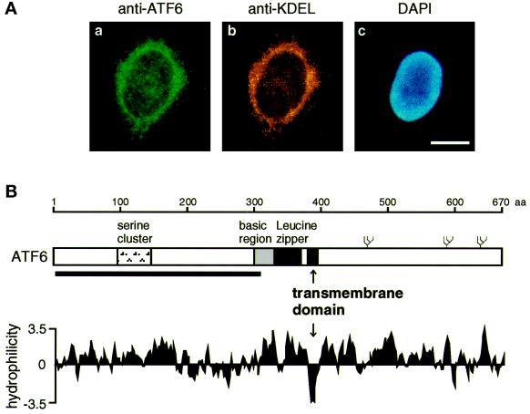
Figure 4.
Identification of an internal hydrophobic stretch that anchors ATF6 in the ER membrane. (A) Indirect immunofluorescence analysis. Unstressed HeLa cells were fixed and stained with anti-ATF6 antibody (a), anti-KDEL antibody (b), or DAPI (c) as described in MATERIALS AND METHODS. Bar, 10 μm. (B) Schematic structure of ATF6 consisting of 670 amino acids. The positions of the serine cluster, basic region, and leucine zipper (Zhu et al., 1997; Yoshida et al., 1998) as well as the transmembrane domain identified in this report are indicated. The thick line below the sequence represents the region (amino acids 6–307) fused with E. coli maltose-binding protein to raise anti-ATF6 antibody (Yoshida et al., 1998). Three potential glycosylation sites are also shown schematically (ψ). The hydropathy index was calculated by the method of Kyte and Doolittle (1982).

