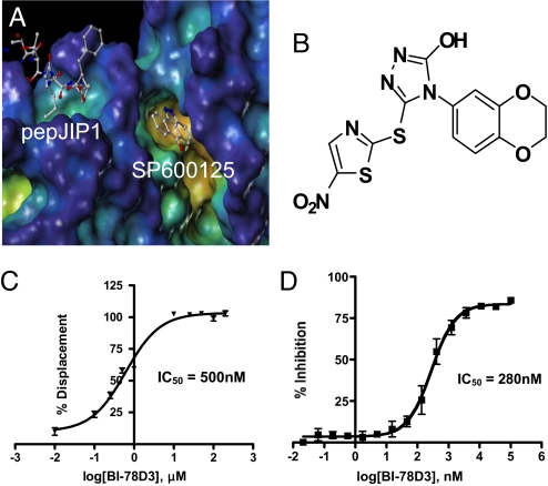
Fig. 1.
In vitro characterization of pepJIP1 and BI-78D3. (A) Surface representation of JNK1 in complex with pepJIP1 (RPKRPTTLNLF) and the ATP mimic SP600125 (PDB-ID 1UKI). Surface generated with MOLCAD (28) and color coded according to cavity depth (blue, shallow; yellow, deep). (B) Chemical structure of BI-78D3. (C) Dose dependent displacement of biotinylated pepJIP1 from GST-JNK1. (D) Kinase inhibition assay for BI-78D3.

