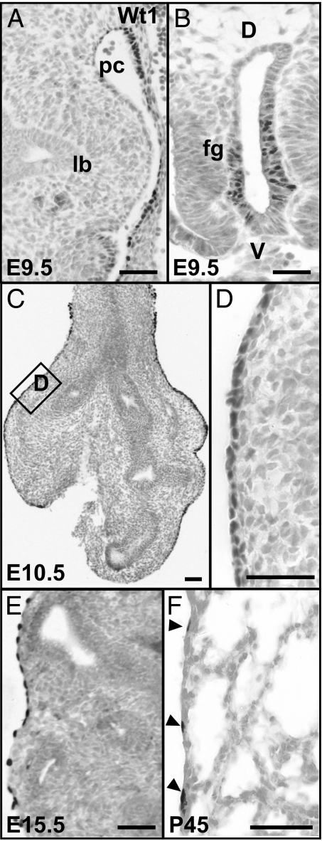
Fig. 1.
Expression of Wt1 protein in the developing and adult lung. (A) At E9.5, Wt1 protein is not detected in the primary lung buds, but in mesothelial cells along the wall of the pericardio-peritoneal cavity. (B) Wt1-positive cells in the ventral epithelium of the unseparated foregut. (C) At E10.5, Wt1-positive mesothelium now covers the surface of both the trachea and lungs. (D) A magnified view of the boxed region in C to show the Wt1-positive and tightly packed mesothelial cells. Wt1-positive mesothelial cells at E15.5 (E) and in the adult (arrowheads) (F). Note that at all times examined no Wt1-positive cells are present either in the epithelium or mesenchyme within the lung; lb, lung bud; pc, pericardio-peritoneal cavity; fg, foregut. (Scale bars, 50 μm.)

