Fig. 2.
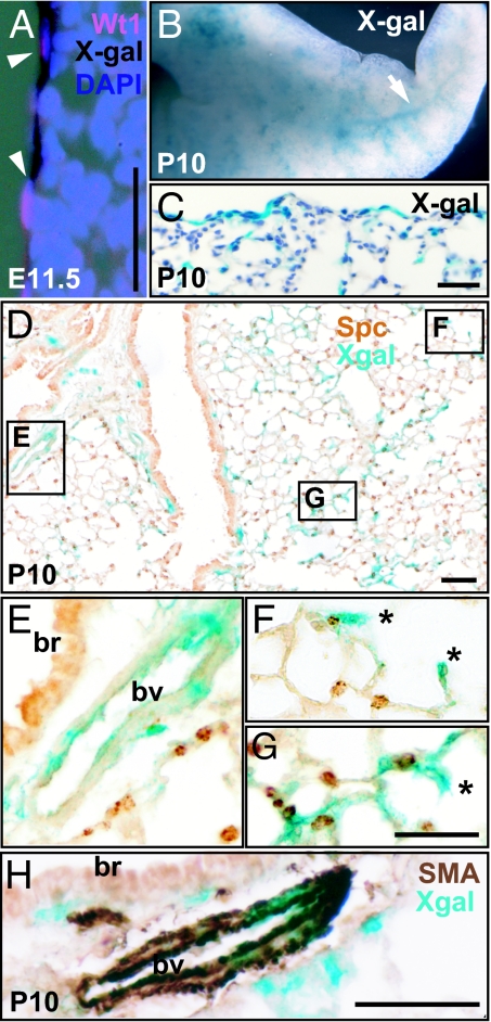
Lineage-labeled mesothelium and its descendents in Wt1-Cre;Rosa26RlacZ lungs. (A) Colocalization of X-Gal staining (black) and Wt1 protein (red) on the surface of E11.5 lung. Nuclei are counterstained with DAPI. (B) Whole mount X-Gal staining of P10 lung. The arrow indicates the network of X-Gal stained cells within the lung. (C and D) Sections after whole-mount X-Gal staining to show that X-Gal positive cells are located both on the surface and within the lung tissue. (D–G) Lung section stained with antibody against SftpC. (E–G) Magnified views of the boxed regions in D. (E) X-Gal positive cells are incorporated into the artery close to the bronchus. (F and G) Two examples of X-Gal cells in alveoli possibly pericyte, endothelial cell, or myofibroblast (indicated by *). (H) Colocalization of alpha-SMA (brown) and X-Gal (blue) in the artery wall next to a bronchus. Note that smooth muscle cells underneath the bronchus do not colocalize with X-Gal; br, bronchus; bv, blood vessel. (Scale bars, 50 μm.)

