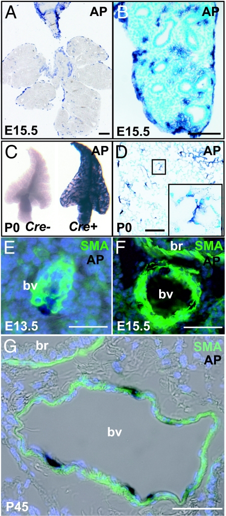
Fig. 3.
Localization of lineage-labeled mesothelial cells and their descendants in Wt1-Cre;Rosa26RCAG-hPLAP lungs. (A and B) Sections of E15.5 lung after whole-mount staining for AP. Note that AP-positive cells are present both on the surface and inside the lung. (C) Whole-mount view of a lobe of AP-stained Wt1-Cre;Rosa26RCAG-hPLAP P0 lung (Left) and Rosa26RCAG-hPLAP control lung, which has no Wt1-Cre transgene (Right). (D) Section of P0 lung after whole-mount AP staining. Inset shows AP-positive cells in the alveoli. (E–G) Colocalization of SMA and AP in some of the vascular smooth muscle cells at E13.5 (E), E15.5 (F), and P45 (G); br, bronchus; bv, blood vessel. [Scale bars: 100 μm (A–D) and 50 μm (E–G).]

