Fig. 4.
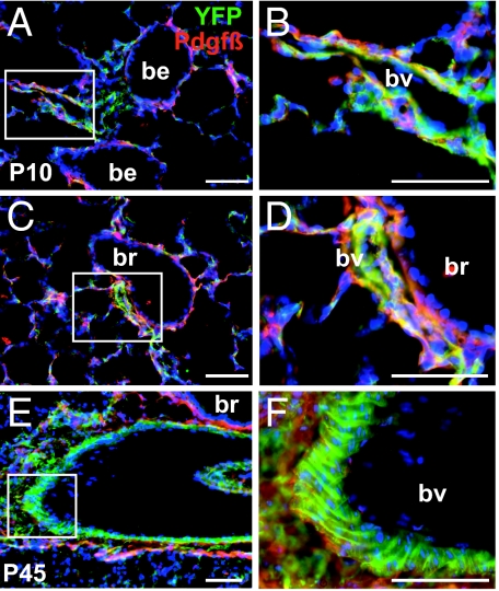
Lineage-labeled cells in Wt1-Cre;Rosa26REYFP lungs. (A–F) Immunohistochemistry of sections of P10 (A–D) and P45 (E–F) lungs from Wt1-Cre;Rosa26REYFP mice with antibody to GFP (green) and PDGF receptor-beta (red). (B, D, and F) Magnified view of the boxed regions in A, C, and E, respectively. Note the presence of lineage-labeled cells that also express PDGF receptor-beta on their surface in the relatively thin walls of vessels closely associated with bronchioles (A–D) and in the thicker walls of a vessel (presumed artery) associated with a larger bronchus (E and F). Note also in F the typical organization of lineage-labeled smooth muscle in the thick mural wall of the presumptive artery. In all sections lineage-labeled cells are absent from the population of airway smooth muscle. All nuclei are counterstained with DAPI; be, bronchiole; br, bronchus; bv, blood vessel. (Scale bars, 50 μm.)

