Figure 2.
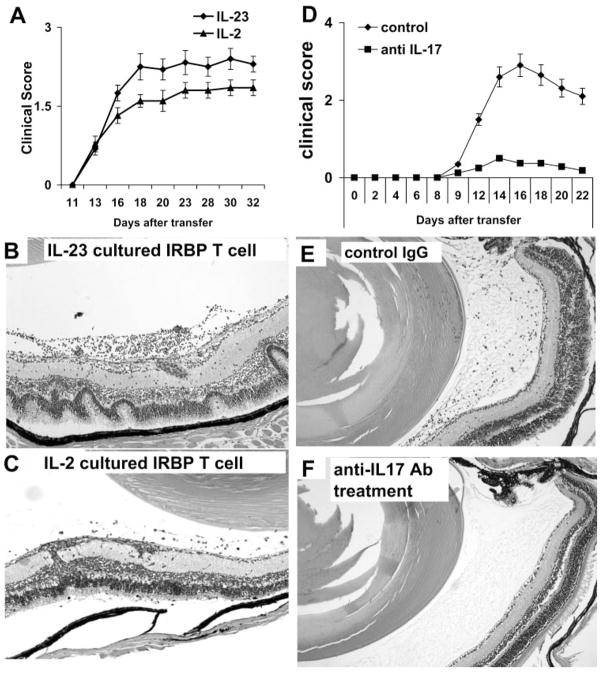
Determination of the uveitogenic activity of IRBP-specific T cells expanded by IL-23. Unfractionated T cells from IRBP1–20–immunized B6 mice at p.i. day 13 were exposed to immunizing peptide in the presence of IL-2 or IL-23 (10 ng/ mL)-containing medium for 3 days. Activated T-cell blasts were separated by Ficoll gradient centrifugation, and 5 × 10⁶ activated T cells were adoptively transferred into a naive B6 mouse. (A) Clinical score by funduscopy with time (six mice per group) and (B, C) pathologic examination of the retinas of mice receiving IL-23–cultured (Th17) (B) or IL-2-cultured (C) IRBP-specific T cells. (D, E) Recipient B6 mice were injected with four doses (100 μg) of anti–IL-17 antibodies twice a week, and control mice were treated with isotype-matched rat antibody. All animals were induced for EAU by injection of 5 × 10⁶ IL-17+ IRBP-specific T cells. Animals were monitored by funduscope examination (D). Pathologic examinations were conducted 15 days after the disease induction (E, F). Mice treated with anti–IL-17 antibodies had significantly milder disease (F).

