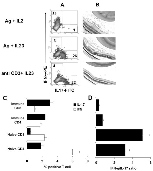
Figure 4.
Comparison of the uveitogenic activity of antigen-specific and nonspecific IL-17+ T cells. (A) Phenotype of the transferred T cells. T cells from IRBP1–20–immunized B6 mice were exposed to the immunizing antigen (10 μg/mL) or to anti-CD3 antibody (1 μg/mL) in the presence of IL-2 or IL-23 (10 ng/mL)-containing medium for 3 days. Activated T-cell blasts were separated by Ficoll gradient centrifugation before adoptive transfer, and the disease-inducing T cells were assessed for expression of IFN-γ and IL-17 by intracellular staining. (B) Disease induction. Cells shown in (A) were adoptively transferred (5 × 10⁶ activated T cells/mouse) to naive B6 mice, and the retinas were examined at p.i. day 15. Antigen/IL-2–expanded cells (top) and antigen/IL-23–expanded cells (center) were pathogenic (infiltration of immune cells and retinal tissues disarranged), whereas anti-CD3 antibody/IL-23–expanded cells were not. (C, D) Expression of IFN-γ and IL-17 by CD4 and CD8 T cells from naive and antigen-immunized B6 mice. Nylon wool–enriched T cells prepared from naive or IRBP–immunized B6 mice and stimulated in vitro with anti-CD3 for 3 days. Separated T-cell blasts were intracellularly stained with anti–IFN-γ and anti–IL-17. Each group consisted of 10 mice. (D) IFN-γ /IL-17 ratio.

