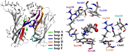
FIGURE 1.
Two adjacent GABAC receptor subunits showing the orientation of GABA in the binding site after the initial minimization of the docked structure in the homology model. The ligand binding site consists of residues from loops A–C of one subunit and loops D–F of the adjacent subunit (left). A closeup of the binding pocket showing the residues referred to in this study (right); the residue labels are colored as the loops they belong to.

