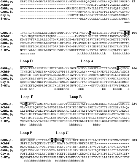
FIGURE 2.
Multiple sequence alignment of representative GABAC, AChBP, GABAA, glycine, and 5-HT3A receptor subunits (GABAC ρ1 receptor subunit numbering). The binding loops A–F are indicated by lines above the alignment. The residues in the binding pocket shown in Fig. 2 are in solid boxes. Based on the secondary structure of AChBP, residues belonging to α-helices, β-sheets, and 310-helices are labeled with a, b, and n, respectively.

