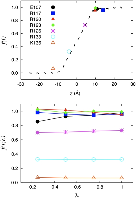
FIGURE 5.
KvAP voltage sensor. Illustration of the charging FEP technique based on Eq. 29 to determine the fraction of the field for specific residues for the fragment of the voltage sensor of the KvAP bacterial channel. (Top) The fraction of the field felt at each of the charged residues is shown with the position of their center of charge along the z axis: Glu107 (10.8 Å); Arg117 (14.1 Å); Arg120 (9.7 Å); Arg123 (10.0 Å); Arg126 (4.4 Å); Arg133 (−4.2 Å); and Lys136 (−12.7 Å). (Bottom) The variation of the fraction of the field extract at different values of the thermodynamic coupling parameter λ during the FEP calculations. The position assigned to each residue along the z axis is based on the geometric center of charge 

