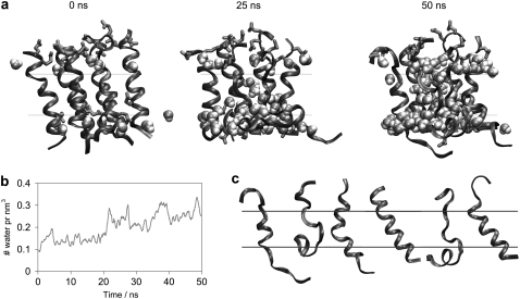
FIGURE 6.
(a) Formation of the leaky membrane by alamethicin peptides. Six alamethicin peptides shown in ribbon with the hydrophilic side chains in licorice. Water in the middle 20 Å of the bilayer and within 8 Å of the six peptides is shown in VDW representation. The middle 12 Å of the bilayer is marked by black lines. (b) Water content of the bilayer. The density of water in the middle 12 Å of the bilayer (calculated at each time step, and subjected to a running average over windows of 0.5 ns) is depicted. (c) Individual conformations of the peptides from the cluster shown in a at t = 50 ns of the AA simulation.

